Legacy: Ultrasound
HOCUS: The Haskins Optically Corrected Ultrasound System
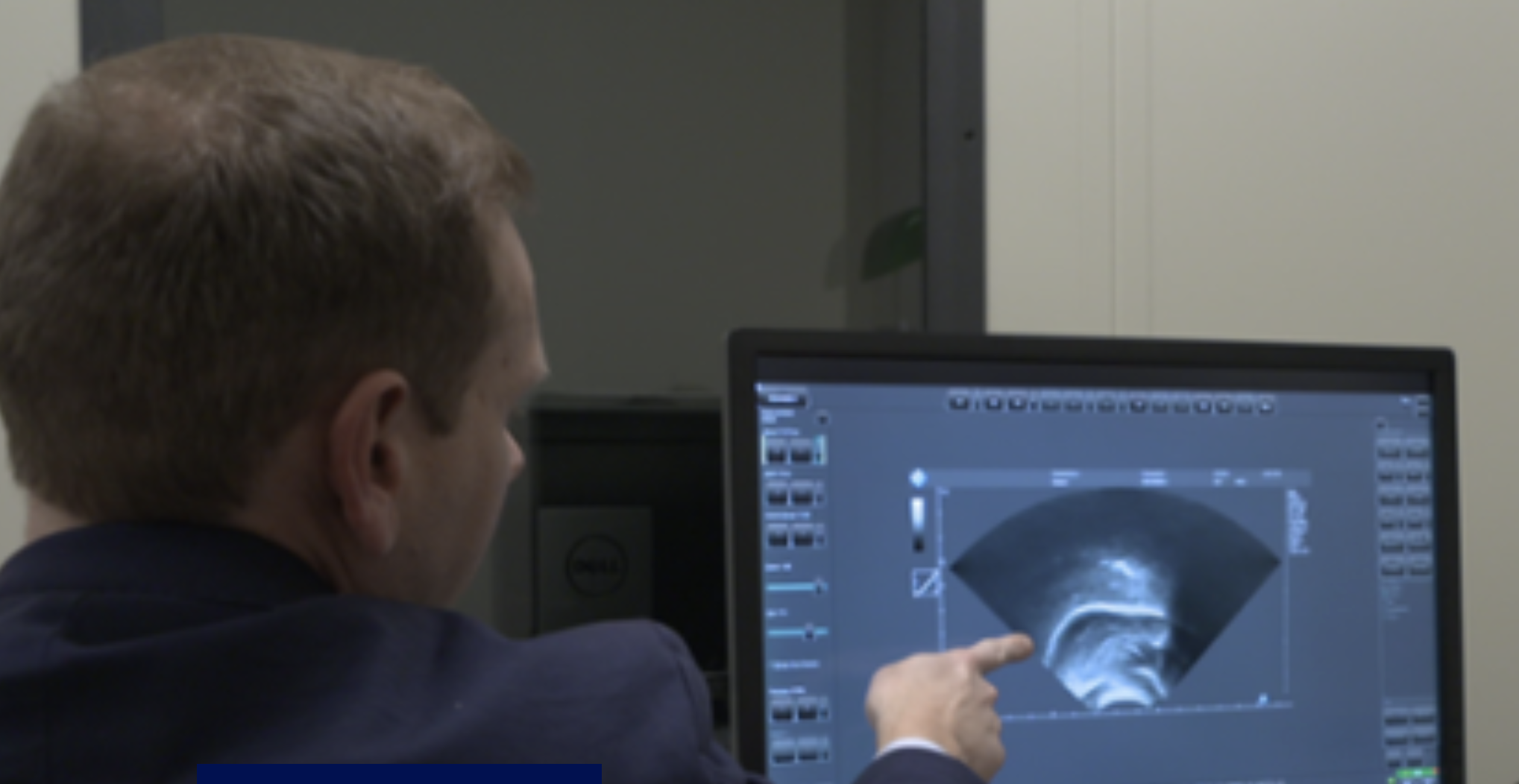
In the early 2000s, Douglas Whalen, Khalil Iskarous, and colleagues pioneered the pairing of ultrasound, used in this case to monitor speech articulators that cannot be seen, and Optotrak, an opto-electronic position-tracking device, used here to monitor visible articulators. The optical system tracks the location of external structures in 3-dimensional space using infrared emitting diodes (IREDs). By tracking 3 or more IREDs on the head and a similar number on an ultrasound transceiver, the transduced image of the tongue can be corrected for the motion of both head and the transceiver and thus be represented relative to the hard structures of the vocal tract. This novel approach provides high-quality, low-cost imaging of most of the tongue surface during fairly unconstrained speech. This work has continued.
Selected References:
Whalen, D.H., Iskarous, K., Tiede, M.K., Ostry, D.J., Lehnert-LeHouillier, H., Vatikiotis-Bateson, E., and Hailey, D.S. (2005). The Haskins Optically Corrected Ultrasound System (HOCUS). Journal of Speech, Language, and Hearing Research, Vol. 48, June 2005, 543-552. (PDF)
Iskarous, K. (2005). Detecting the edge of the tongue: A tutorial. Clinical Linguistics & Phonetics, Vol. 19 (6/7), 555-565. (PDF)
Noiray A., Ménard, L., and Iskarous, K. (2013). The Development of Motor Synergies in Children: Ultrasound and Acoustic Measurements. Journal of Acoustical Society of America, Vol. 133 (1), 444-452.
Roon, Kevin D., Kang, Jaekoo, and Whalen, D. H. (2020). Effects of Ultrasound Familiarization on Production and Perception of Nonnative Contrasts. Phonetica, Vol. 77, 350-393.

Ultrasound Visual Feedback
Within the past decade, there has been interest in the clinical application of ultrasound to individuals with speech disorders. By holding an ultrasound transducer under the chin, real-time images of the tongue can be observed. These images can be used to provide both the client and the clinician with information about tongue position and configurations during production of certain speech sounds or during sequences of sounds. Speech-language pathologists can use this information to provide cues to their clients about how to change the tongue position or shape. Ultrasound imaging of the tongue can be used to address articulation of sounds such as /t, d, n, s, z, l, r, k, g/ and vowels.
For a number of years, a group at Haskins Laboratories led by Dr. Jonathan Preston provided some clinical training for speech-language pathologists. This group is no longer active at Haskins. Jonathan, a Haskins affiliate, moved to Syracuse University where he continues working on visual feedback treatments for speech, including ultrasound imaging of the tongue and visual acoustic biofeedback. For a complete listing of Dr. Preston's research go to his Laboratory Website .
Selected References:
Jonathan L. Preston, Tara McAllister, Emily Phillips, Suzanne Boyce, Mark Tiede, Jackie Sihyun Kim, and Douglas H. Whalen (2019). Remediating Residual Rhotic Errors With Traditional and Ultrasound-Enhanced Treatment: A Single-Case Experimental Study. American Journal of Speech-Language Pathology, Vol. 28, 1167-1183.
Laine Cialdella, Heather Kabakoff, Jonathan Preston, Sarah Dugan, Caroline Spencer, Suzanne Boyce, Mark Tiede, D. Whalen, and Tara McAllister (2021). Auditory-perceptual acuity in rhotic misarticulation: baseline characteristics and treatment response, Clinical Linguistics & Phonetics, Vol. 35, issue 1.